Author: mmanning
-
Dapeng Bi accepts tenure track position at Northeastern
Former Manning group postdoc Dapeng (Max) Bi has accepted a tenure track position at Northeastern University beginning in 2017. He is currently a postdoctoral fellow at Rockefeller University. Congratulations, Max! His website is here.
-
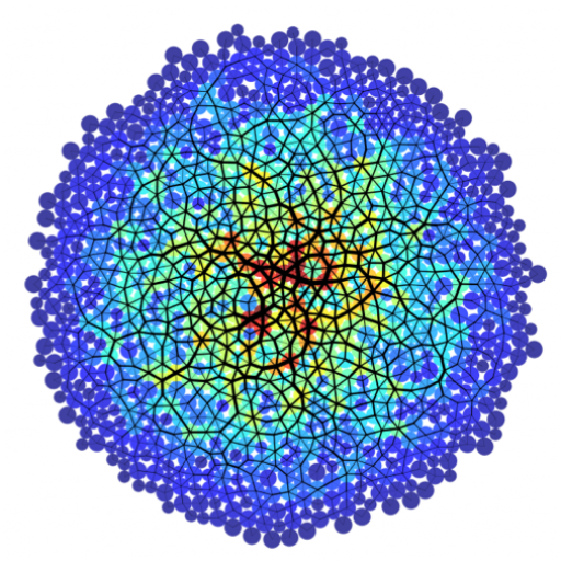
Manuscript published in PRX!
Our manuscript in collaboration with the Marchetti group describing a glass transition in a new self-propelled voronoi model for active tissues was just accepted at PRX. Congratulations, Max and Xingbo! Dapeng Bi, Xingbo Yang, M. Cristina Marchetti, M. Lisa Manning. Motility-driven glass and jamming transitions in biological tissues. PRX 6 021011 2016. http://arxiv.org/abs/1509.06578
-
Physics of Development and Disease
Manning is co-organizing the Aspen Center for Physics Workshop, Physics of Development and Disease, March 27-April 1 2016 in Aspen, CO. The website for the conference is here. Manning is giving a public lecture, and Merkel is presenting a talk entitled, “Glassy dynamics in a model for confluent three-dimensional tissues”.
-
Manning wins IUPAP Young Investigator Prize
It was announced this week that Lisa Manning has won the 2016 IUPAP Young Investigator Award given by the C3 (Statistical Physics) commission, along with Martin Lenz. The award is given “in recognition of her outstanding statistical physics contributions to the fields of granular materials, jamming, and biological cell dynamics.” The award is given to…
-
Manning group publishes in Nature Physics
Our manuscript “A density-independent rigidity transition in biological tissues” with Max Dapeng Bi as lead author and Lisa Manning as corresponding author, was published online this week in Nature Physics! Congrats Max! Nature Physics online 21 Sept 2015
-
Manning elected as Soft Matter GRC vice chair, gives invited talk
Lisa gave an invited talk at the 2015 Soft Matter Gordon research conference in New London, NH, and was elected vice-chair of the conference in 2017, to serve as chair in 2019. A news article on the wonderful representation of SU soft matter at the conference is here: http://asnews.syr.edu/newsevents_2015/releases/physics_grc_conference.html
-
Manning group receives NIH R01 funding
Lisa is the PI on a recently awarded R01 proposal to the NIH, “Quantitative Modeling of Cell Shape Changes During Organogenesis”. The award is for $1.02 million over 4 years, to be shared with collaborator Jeff Amack at Upstate Medical University. The award abstract reads: During embryonic development, the proper formation of tissues and organs…
-
Manning group publishes in Nature Materials
Lisa and Max worked together with members of Jeff Fredberg’s group at the Harvard School of public health to see if their theoretical predictions could be verified in human cells from asthma and non-asthma patients. The theoretical results were spectacularly confirmed, as discussed in a paper published online last week in Nature Materials: http://www.nature.com/nmat/journal/vaop/ncurrent/full/nmat4357.html A…
-
Manning group featured in NSF discoveries
A nice article (with a strange titles) about our group’s research was recently highlighted on the NSF “Discoveries” page: http://www.nsf.gov/discoveries/disc_summ.jsp?cntn_id=134489
-
Manning group wins SCIALOG award
SU news article on the award: http://news.syr.edu/physicist-awarded-grant-to-study-physical-cell-biology-93769/ As we grow from a fertilized egg into a human being, our cells push and pull on one another, shaping our tissues, our organs, our bones, and our bodies. Unfortunately we don’t know much about how these microscopic forces, both within and between cells, allow large multi-cellular structures…