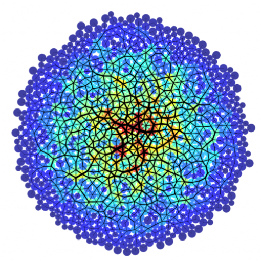
During embryonic development individual cells must actively change their shapes in a coordinated way to generate large-scale patterns that are critical for the formation of tissues and organs. An open question is how these programmed cell shape changes are regulated via a combination of mechanical forces and biochemical signaling pathways. In order to make progress in understanding and treating developmental diseases, we must account for both types of interactions and use predictive models to identify feedbacks between them.
The zebrafish embryo is an excellent vertebrate system for investigating tissue patterning and organogenesis. To study cell shape changes during organogenesis, we are using the zebrafish organ of asymmetry—called Kupffer’s vesicle (KV)—as a model organ. KV is a simple organ with a fluid-filled lumen surrounded by a single layer of monociliated epithelial cells. Motile cilia projecting into the lumen create an asymmetric fluid flow that is necessary for specifying the left-right body axis. We have identified cell shape changes during KV morphogenesis that are essential for KV organ formation and function. This morphogenetic process, which we refer to as ‘KV remodeling,’ provides an opportunity to characterize biophysical mechanisms that drive organogenesis.
Manuscript: Regional Cell Shape Changes Control Form and Function of Kupffer’s Vesicle in the Zebrafish Embryo
Currently, Craig Fox is working in collaboration with Amack lab to image and analyze fluid flows inside the KV. He is also working to understand how the mechanical properties of tissues like the notochord that are near the KV influence cell shapes and left-right patterning.
